
Interventional Radiology Treatment of Liver Cancer
Surgical removal of liver tumors offers the best chance for a cure. Unfortunately, liver tumors are often inoperable because the tumor may be too large, or has grown into major blood vessels or other vital structures. Sometimes, many small tumors are spread throughout the liver, making surgery too risky or impractical.
- Surgical removal is not possible for more than two-thirds of primary liver cancer patients and 90 percent of patients with secondary liver cancer.
- cancers that primarily arise from the liver cells themselves
- those that spread or metastasize to the liver from other sites such as the colon, lung, or breast.
Historically, chemotherapy drugs have been generally ineffective at curing liver cancer.
- Surgical removal is not possible for more than two-thirds of primary liver cancer patients and 90 percent of patients with secondary liver cancer.
There are generally two types of cancer that involve the liver:
- cancers that primarily arise from the liver cells themselves
- those that spread or metastasize to the liver from other sites such as the colon, lung, or breast.
Historically, chemotherapy drugs have been generally ineffective at curing liver cancer.
Primary Liver Cancer
The most common form of primary liver cancer is hepatocellular carcinoma (HCC).
This is a tumor that begins in the main cells of the liver (hepatocytes). HCC most frequently occurs in those who have a form of liver disease called cirrhosis.
Cirrhosis occurs when the liver becomes diseased and develops scarring, usually over a period of years. The liver attempts to repair or regenerate itself. This process can lead to the formation of tumors. In the United States, the most common causes of cirrhosis are alcohol abuse and chronic infection with the liver virus hepatitis B or C.
The incidence of primary hepatocellular carcinoma is on the rise worldwide because of the increase of hepatitis C.
About 18,500 cases of primary liver cancer are diagnosed each year.
Primary liver cancer is twice as common in men as in women.
About 18,500 cases of primary liver cancer are diagnosed each year.
Primary liver cancer is twice as common in men as in women.
Metastatic Liver Cancer
Cancer may spread from any part of the body to the liver. There the cancer cells may grow for months or years before they are detected. One of the most common sources of metastatic liver cancer is from tumors of the colon and rectum.
Patients with other types of cancer also are at risk for liver cancer. The liver serves as a way-station for cancer cells that circulate through the bloodstream. These cells may grow and form tumors in the liver. It is estimated that as many as 70 percent of all people with uncontrolled cancer will eventually develop secondary liver tumors, or metastases (tumors formed by primary cancer cells that have spread from other cancer sites).
140,000 people in the United States are diagnosed with colon cancer each year
Roughly 1/2 of these patients will develop tumors in their liver
1 in 10 of these patients will have a chance for a cure by having the liver tumors removed surgically.
140,000 people in the United States are diagnosed with colon cancer each year
Roughly 1/2 of these patients will develop tumors in their liver
1 in 10 of these patients will have a chance for a cure by having the liver tumors removed surgically.

Radiofrequency Ablation (RFA)
RFA is a therapy that involves placement of a small needle through the skin into the tumor and killing the cancer by use of thermal energy or heat. The cancer cells are essentially “cooked.”
The needles are guided into place by either CT or ultrasound. This technique can be used for both primary liver cancers (hepatocellular carcinoma or HCC) and metastatic disease. In general, the best results are achieved with single tumors, one that are smaller (less than 3 cm), and those that are primary liver cancers.
RFA is usually performed as an outpatient with conscious sedation or general anesthesia. Complications occur less than 2% of the time.
Selection criteria for RF ablation in the liver:
- Four or fewer primary/secondary malignant hepatic tumors
- Tumors less than 5 cm (about 2 inches)
- Patient not a candidate for liver surgery
- Life expectancy of more than six months
- No bleeding issues
- No active liver infection.
Embolization
Embolization is a well established interventional radiology technique that is used to treat trauma victims with massive bleeding, to control hemorrhage after childbirth, to decrease blood loss prior to surgery, and to treat tumors.
In treating cancer patients, interventional radiologists use embolization to cut off the blood supply to the tumor (embolization), deliver radiation to a tumor (radioembolization), or combine this technique with chemotherapy to deliver the cancer drug directly to the tumor (chemoembolization).
As vascular experts, interventional radiologists are uniquely skilled in using the vascular system to deliver targeted treatments via catheter throughout the body. In treating cancer patients, interventional radiologists can attack the cancer tumor from inside the body without medicating or affecting other parts of the body.
Tumors need a blood supply, which they actively generate, to feed themselves and grow. As vascular experts, interventional radiologists are uniquely skilled in using the vascular system to deliver targeted treatments via catheter throughout the body. In treating cancer patients, interventional radiologists can attack the cancer cells by directly injecting substances into their blood supply.
Radioembolization (Yttrium Y-90)
Radioembolization is a combination of radiation therapy and embolization. Tiny glass or resin beads are injected through a small tube or catheter placed into the liver artery from a tiny skin nick in the groin. These tiny beads or microspheres become lodged or trapped in the small vessels of the tumor where they deliver high radiation dose directly to the cancer cells. These beads or microspheres are filled with a radioactive isotope Yttrium Y-90. This is a palliative treatment, meaning it does not offer a cure but instead helps slow down the progression of disease and alleviate symptoms. Life can be extended months to years and improve symptoms as well as quality of life. The radiation dose is delivered to the tumor over the next 10 to 14 days. The radiation disappears after about 30 days.
The radioembolization procedure is a two-step process. An initial angiogram is performed one to two weeks prior to injection of the Yttrium Y-90 microspheres. This enables the interventional radiologist to “map out” the arterial anatomy and to block off several small arteries to prevent the microspheres from going to places other than the tumor. Complications from radioembolization are about 1 in 20 patients. A few patients experience some side effects called post-embolization syndrome, including nausea, vomiting, and fever. Pain is the most common side effect that occurs because the blood supply to the tumor is cut off. This can be controlled by medication. You should be able to resume normal activities one to two days after the procedure.
Chemoembolization
This procedure is similar to that described above for radioembolization except certain chemotherapy drugs are injected into the arteries supplying the tumor rather than the radioactive microspheres. Our interventional radiologists will work with you and your oncologist to determine which therapy is right for you.
Selected Portal Vein Embolization
VIRA also offers additional specialized therapies. Sometimes your surgeon may plan to remove a portion of your liver that contains the tumor but the remaining part would be too small to adequately support your liver function. In these cases, we can do a specialized presurgical embolization of a vein in your liver (portal vein) that can allow the healthy part of your liver to grow or hypertrophy over several months prior to your surgery and therefore enable the surgeon to remove the diseased portion.
We welcome the opportunity to work with you and your oncologist to provide minimally invasive treatments to prolong survival and enhance your quality of life.

Cancer Resources

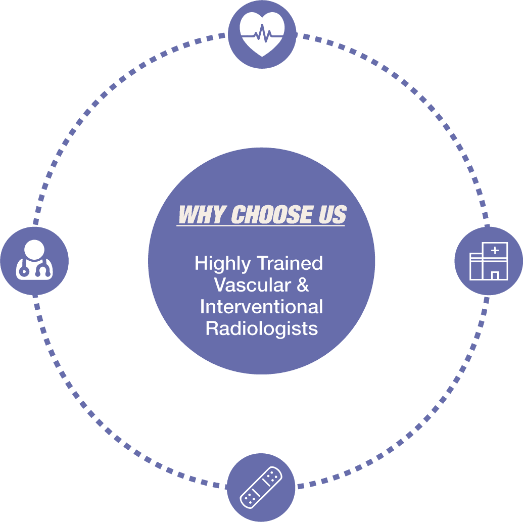
Highly Trained InterventionalRadiologists & Expert Staff
Highly Trained Interventional
Radiologists & Expert Staff
Fellowship-trained, Board-Certified doctors who are experts in noninvasive therapies. Friendly knowledgeable staff who put your experience and comfort first.
Thorough Diagnosis & Personalized Care
State of the Art Facility & Treatment Options
Innovative minimally invasive procedures performed in an office-based setting provide a more personal, comfortable and private experience than a hospital or surgical center.
Minimally Invasive Procedures
Quicker recovery, faster healing,
and little-to-no scarring.